Lumbosacral Plexopathy

Lumbosacral plexopathy is an injury to or involvement of one or more nerves that combine to form or branch from the lumbosacral plexus. This involvement is distal to the root level.

shutterstock 1865161186

Lumbosacral Plexopathy Summary

Lumbosacral plexopathies (LSP) represent a distinct group of disorders of the peripheral nervous system due in part to their relative rarity in comparison to other peripheral nerve disorders and also due to their wide array of etiologies. Initial treatment is based on both the presenting symptoms and the cause of the lumbosacral plexopathy. Some patients may benefit from injection treatments including psoas compartment block and botox injections.

Procedures offered for Lumbosacral Plexopathy

inguinal nerve opt 1

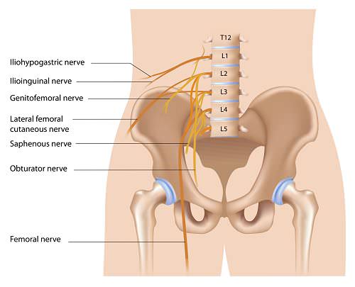
Anatomy Of Lumbar Plexus

The lumbar plexus originates from the first, second, third, and fourth lumbar nerves. The fourth lumbar nerve makes a contribution to both the lumbar and the sacral plexus. There is typically a small communication from the twelfth thoracic nerve as well. These nerve roots divide into the dorsal rami and the ventral rami as they exit through the intervertebral foramina. The dorsal or posterior rami innervate the paraspinal muscles and supply nearby cutaneous sensation. The ventral or anterior rami of the lumbar plexus form the motor and sensory nerves to the anterior and medial sides of the thigh and the sensation on the medial aspect of the leg and foot. The undivided anterior primary rami of the lumbar and sacral nerves also carry postganglionic sympathetic fibers that are mainly responsible for vasoregulation of the lower extremities. The branches of the lumbar plexus include the iliohypogastric, ilioinguinal, genitofemoral, femoral, lateral femoral cutaneous and obturator nerves. The lumbar portion of the plexus lies just anterior to the psoas muscle.

Aetiology

Lumbosacral plexopathy has been recognized as a clinical entity or complication in a variety of surgical procedures, trauma, and obstetric surgery or delivery and as a clinical finding or sequela in treatment of pelvic tumors.

Trauma

Traumatic pelvic fractures have a 30% incidence of lumbosacral plexus injury. Sacral fractures have become recognized as an essential consideration in pelvic trauma because of their high association with lumbosacral nerve deficits. This can have a profound influence on prognosis and level of functional recovery. Trauma is a common cause of retroperitoneal hemorrhage, which can injure the lumbosacral plexus.

Postpartum Plexopathy

Compression injury to the lumbosacral plexus during labor and delivery, known as postpartum lumbosacral plexopathy, is underappreciated and often misdiagnosed. Although most large series place the incidence of this disorder at one in 2600 births, there are likely many milder cases that never reach medical attention.

The mechanism of injury likely involves compression of the fetal head against the underlying pelvis and lumbosacral plexus. Postpartum lumbosacral plexopathy results primarily from compression of the lumbosacral trunk. These are the fibers from the L4 and L5 roots, which join together to descend into the pelvis to reach the sacral plexus. When the lumbosacral trunk crosses the pelvic outlet, the fibers lie exposed (no longer protected by the psoas muscle) as they rest against the sacral ala near the sacroiliac joints. At this point, the fibers are most exposed and susceptible to compression. The origin of the superior gluteal nerve lies close by and may also be compressed. The fibers that eventually form the peroneal division of the sciatic nerve lie posteriorly, closest to the bone, and are more vulnerable to compression than the tibial division fibers. Accordingly, peroneal fibers are often most affected, with some women presenting with a postpartum footdrop, not infrequently misdiagnosed as peroneal palsy at the fibular neck.

Weakness may be noticed immediately or within the first few days after delivery. In addition to peroneal weakness, examination often shows mild weakness of knee flexion (hamstrings) and hip abduction, extension, and internal rotation (glutei, tensor fascia latae), demonstrating that the lesion is clearly beyond the peroneal territory. Sensory disturbance is most marked over the dorsum of the foot and lateral calf but may be patchy and involve the sole of the foot, posterior calf, and thigh.

Several factors predispose to this injury, including a first pregnancy, a large fetal head with a small maternal pelvis (cephalopelvic disproportion), a small mother (less than 5 feet in height), and a prolonged or difficult labor. Although rare patients may be left with permanent weakness, the prognosis is excellent in most cases. The presumed mechanism of injury involves compression that leads to ischemia and mechanical deformation of nerve fibers, which in turn lead to demyelination and, if severe enough, axonal loss. There is no tearing, shearing, or disruption of basement membranes. Thus, even in cases with severe axonal loss, recovery often is complete.

Inflammatory (idiopathic Lumbosacral) Plexitis

Idiopathic plexitis occurs in the lumbosacral plexus, although it is far less frequent than its upper extremity counterpart, brachial neuritis (now most properly referred to as neuralgic amyotrophy). The underlying pathology is not completely known, although it is probably inflammatory, often occurring within a few weeks of a possible inciting immunologic event, such as a cold, flu, or immunization. In some cases, there is no clear inciting event. Patients initially develop severe deep pain, either proximal in the pelvis or in the upper leg. Although the pain characteristically persists for 1 to 2 weeks, as in idiopathic brachial plexitis, in some patients pain may become a disabling symptom, lasting many months. Because both the upper and lower plexus may be involved, many different patterns of weakness and sensory loss may develop.

The classic presentation is that of acute, severe pain that subsides over several weeks, followed by weakness that recovers over many months to years. In contrast to these monophasic presentations, other patients may present with a progressive course. Some patients have been described with a progressive, painful lumbosacral plexopathy, often with an elevated sedimentation rate, who have improved with steroids or other immunosuppressives. Such cases may represent localized forms of vasculitic neuropathy.

Iatrogenic

Gynecologic surgery is thought to be one of the most common causes of femoral nerve injury and lumbosacral plexus nerve injuries. Abdominal hysterectomy is the surgical procedure that has been most frequently implicated. The mechanisms of neurologic injury that have been established include improper placement or positioning of self-retaining or fixed retractors, incorrect positioning of the patient in lithotomy position preoperatively or prolonged lithotomy positioning without repositioning, and radical surgical dissection resulting in autonomic nerve disruption.

Lumbosacral injury can also occur after appendectomy and inguinal herniorrhaphy. Patients who are thin, diabetic, or elderly are at increased risk for such an injury. Injury to the lumbosacral plexus with clinical findings occurs in up to 10% of hip replacement procedures, and injury that is subclinical but detected electromyographically occurred in up to 70% of patients.

Retroperitoneal hemorrhage causing lumbosacral plexopathy may occur with no precipitating injury, especially in patients on anticoagulant therapy. Retroperitoneal hematoma can also occur in patients undergoing femoral vessel catheterization for stents and dialysis.

Tumors

Both pelvic malignant neoplasms and treatment of pelvic tumors can damage the lumbosacral plexus. Lumbosacral radiculopathy is most common with gynecologic tumors, sarcomas, and lymphomas. Neoplastic plexopathy is characterized by severe and unrelenting pain, typically followed by weakness and sensory disturbances. Pelvic radiation therapy may cause a delayed lumbosacral plexopathy that can occur months to years after completion of treatment; the median amount of time from the completion of treatment to onset of symptoms is about 5 years. Chemotherapeutic agents can also cause symptoms of lumbosacral radiculopathy. Cisplatin, 5-fluorouracil, mitomycin C, and bleomycin have been implicated in the majority of these plexopathies.

Metastatic or tumor extension into the lumbosacral plexus and malignant psoas syndrome have been described in the literature. It is characterized by proximal lumbosacral plexopathy, painful fixed flexion of the ipsilateral hip, and radiologic or pathologic evidence of ipsilateral psoas major muscle malignant involvement.

Diabetic Amyotrophy

Diabetic lumbosacral radiculoplexus neuropathy is a subacute, painful asymmetric lower limb neuropathy that is associated with significant weight loss (at least 10 pounds), type 2 diabetes mellitus, and relatively recent diagnosis of diabetes with relatively good glucose control. The underlying pathophysiologic mechanism is thought to be immune mediated with microvasculitis of the nerve rather than a metabolic issue caused by diabetes; nondiabetic lumbosacral radiculoplexus neuropathy has also been documented with similar clinical and pathophysiologic features.

Vascular

Vascular causes of lumbosacral plexopathies may include diabetic amyotrophy and connective tissue diseases that may be associated with vasculitis, such as systemic lupus erythematosus, rheumatoid arthritis, and polyarteritis nodosa.

Clinical Presentation

Plexopathies may vary considerably in their presentation, depending on the location and degree of involvement. Lumbosacral plexopathy often begins with leg pain radiating to the low back and buttocks and progressing posterolaterally down the leg, soon followed by symptoms of numbness and weakness. Lumbosacral plexus injuries are often associated with a footdrop and sensory changes to the top of the foot.

Lumbosacral plexus lesions usually are divided clinically into those affecting the upper lumbar plexus and those affecting the lower lumbosacral plexus, analogous to the underlying anatomic division. Lumbar plexopathies affect predominantly the L2–L4 nerve fibers, resulting in weakness of the quadriceps, iliopsoas, and hip adductor muscles (femoral and obturator nerves). The knee jerk is frequently depressed or absent. Pain, if present, usually is located in the pelvis with radiation into the anterior thigh. Sensory loss and paresthesias occur over the lateral, anterior, and medial thigh and may extend down the medial calf.

Lesions of the lower lumbosacral plexus predominantly affect the L4–S3 nerve fibers. Patients describe a deep boring pain in the pelvis that can radiate posteriorly into the thigh with extension into the posterior and lateral calf. The ankle jerk may be depressed or absent. Sensory symptoms and signs may be seen over the posterior thigh and posterior-lateral calf and in the foot. Accordingly, patients may present with footdrop and sensory disturbance over the dorsum of the foot and lateral calf.

Diabetic plexopathy typically starts as an identifiable onset of asymmetric lower extremity symptoms that most typically involve the thigh and hip with pain that progresses to include weakness, which then becomes the main disabling symptom. In a few months, this usually evolves into bilateral symmetric weakness and pain with distal as well as proximal involvement. Although motor findings are prominent, sensory and autonomic nerves have also been shown to be involved.

Bowel and bladder symptoms and sexual dysfunction can occur with involvement of sacral nerve roots.

Patients frequently present with some type of difficulty with mobility and ambulation. Activities such as transferring from one surface to another, rising from a chair, ambulation, grooming, bathing, dressing, and cooking may potentially be affected.

Examination Findings

Clinical examination should include moto testing, sensory testing, muscle stretch reflexes, tone and bowel and bladder function. The pattern of sensory loss, asymmetric reflexes, or weakness is suggestive of multiple nerve or root level involvement. It is important to differentiate a suspected plexus injury from single root level involvement, suggesting a radiculopathy, or more generalized nerve changes consistent with peripheral neuropathy. Edema or swelling in one lower extremity may be suggestive of a pelvic mass or lumbosacral plexus involvement rather than a more global peripheral neuropathy or possible retroperitoneal hematoma or pelvic malignant neoplasm.

Investigations

Electrodiagnostic Testing

The electrodiagnostic evaluation of lumbosacral plexopathy is one of the most effective tools available for differentiation of a specific pattern and severity of nerve involvement. Sensory involvement without motor involvement suggests a lesion distal to the dorsal root ganglion.

The needle electromyographic examination is likely to be the most useful electrodiagnostic technique. Careful examination of proximal and distal musculature demonstrates a pattern of muscle membrane instability in more than one peripheral nerve from different root levels without involvement of the paraspinal muscles.

The sensory nerve conduction studies are crucial in identifying a plexus lesion. Both superficial peroneal and sural sensory studies should be performed in a suspected lower lumbosacral plexopathy, and saphenous studies should be done for a suspected lumbar plexopathy. Sensory nerve action potential (SNAP) amplitudes should be carefully compared from side to side. Decreased SNAP amplitudes generally imply a lesion either at the dorsal root ganglion or distally in the plexus or peripheral nerves, but not at the nerve roots.

Imaging studies

MRI scans and CT scans should be considered if a structural mass is suspected in the pelvic region.

Differential diagnosis

  • Spinal cord injury
  • Cauda equina injury
  • Lumbosacral radiculopathy
  • Multiple peripheral nerve injuries
  • Anterior horn cell diseases
  • Myopathies

Treatment Of Lumbosacral Plexopathy

Initial treatment is based on both the presenting symptoms and the cause of the lumbosacral plexopathy. For example, many obstetric lumbosacral plexus symptoms are treated conservatively. Pelvic masses or a retroperitoneal hemorrhage may require surgical or medical intervention. Neoplastic or radiation-based plexopathy symptoms may need specific medical management, chemotherapy or possibly surgery.

Rehabilitation

Rehabilitation aims to maximize mobility and functional independence. The goals of rehabilitation are preservation of joint range of motion and flexibility, joint protection and pain management.

Pharmacological agents

Antineuropathic drugs including tricyclics, Gabapentanoids and SNRI’s form the basis of pain management in lumbosacral plexopathy.

Immune therapy

Various immunomodulation therapies for diabetic amyotrophy, including corticosteroids, cyclophosphamide, intravenous immune globulin, and plasmapheresis, have been described in a number of case series, most of which report positive outcomes with regard to resolution of pain and weakness. However there are no randomized controlled trials to definitively recommend the use of immunotherapy for diabetic lumbosacral radiculoplexus neuropathies.

Interventional Treatments

Psoas compartment block

Psoas compartment block under ultrasound guidance can help with pain management in lumbosacral plexopathy.

Ultrasonographic visualization of the psoas muscle in adults requires a low frequency transducer (5–8 MHz) due to the depth of the lumbar plexus (5–8 cm). The longitudinal sonographic pattern of the psoas muscle demonstrates a hypoechoic background interspersed with hyperechoic bands (dots on transverse view) representing fibrous structures (or possibly neural elements of the plexus) within the muscle. The kidneys are visualized as oval shaped structures usually at the level of L2 or L3, and therefore can be avoided during ultrasound-guided psoas compartment block. The kidneys can also be seen to move with respiration. The more hyperechoic, wedge-shaped, psoas muscle lies medial and deeper to the kidneys.

Botulinum toxin injection

Botulinum toxin injection has been described for management of psoas syndrome.

Have a question about Lumbosacral Plexopathy?
Or would like to book in with us?

0117 2872383

Get In Touch
murli

Dr Murli Krishna

Consultant Pain Medicine